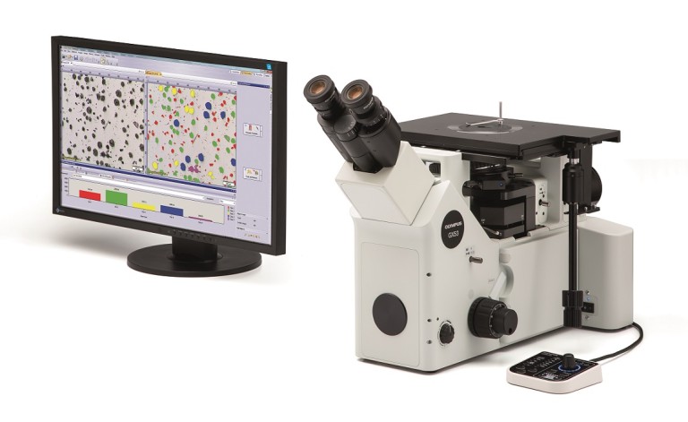
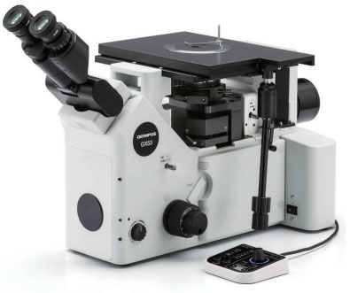
The GX53 inverted microscope is used for a wide range of applications often seen in the steel, automotive, electronics, and other manufacturing industries. The microscope enables users to inspect polished metals and cross-section samples simply by placing them upside down on the stage. The sample does not need to be leveled and can be thick, large, or heavy. The GX53 delivers crisp images that can be difficult to capture using conventional microscopy observation methods. When combined with OLYMPUS Stream image analysis software, the microscope streamlines the inspection process from observation to image analysis and reporting.
Fast Inspections, Advanced Functionality
The GX53 microscopeвҖҷs various observation capabilities provide clear, sharp images, so users can reliably detect defects in their samples. OLYMPUS Stream image analysis software's new illumination techniques and image acquisition options give users more choices for evaluating their samples and documenting their findings.
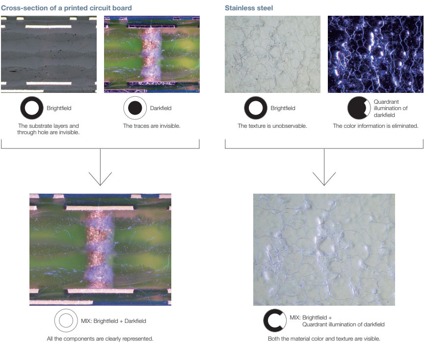
The Invisible Becomes Visible: MIX Technology
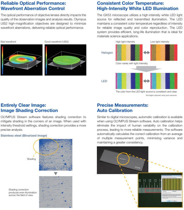
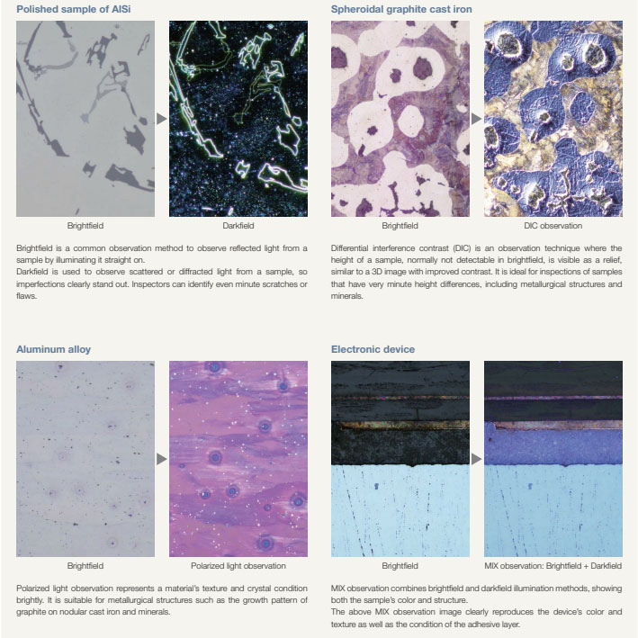
MIX technology produces unique observation images by combining darkfield with another observation method, such as brightfield or polarization. MIX observation enables users to view samples that are difficult to see with conventional microscopes, and represents even small height differences of sample surfaces. The circular LED illuminator used for darkfield observation has a directional darkfield function where one or more quadrants are illuminated at a given time. This reduces a sampleвҖҷs halation and is useful for visualizing its surface texture.
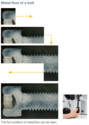
Easily Create Panoramic Images: Instant MIA
With multiple image alignment (MIA), users can stitch images together quickly and simply by moving the XY knobs on the manual stageвҖ”a motorized stage is optional. OLYMPUS Stream software uses pattern recognition to generate a panoramic image, which is suitable for inspections of carburizing and metal-flow conditions.
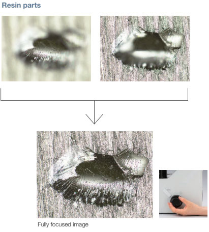
Create All-in-Focus Images: EFI
OLYMPUS Stream softwareвҖҷs extended focus imaging (EFI) function captures images of samples whose height extends beyond the depth of focus. EFI stacks these images together to create a single all-in-focus image of the sample. Even when analyzing a cross-section sample with an uneven surface, EFI creates fully-focused images. EFI works with either a manual or motorized Z-axis and creates a height map to easily visualize structures.
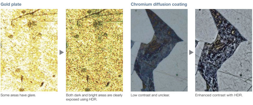
Capture Both Bright and Dark Areas Using HDR
Using advanced image processing, high dynamic range (HDR) adjusts for differences in brightness within an image to reduce glare. It also helps boost the contrast in low-contrast images. HDR can be used to observe minute structures in electric devices and identify metallic grain boundaries.
OLYMPUS Stream Software вҖ“ Optimized for Materials Science
Material inspection, measurement, and analysis are required to comply with industrial standards as well as internal operating procedures. Together, the GX53 microscope and OLYMPUS Stream software support metallurgical analysis methods that comply with different industrial standards. With step-by-step operator guidance, users can analyze their samples quickly and easily.
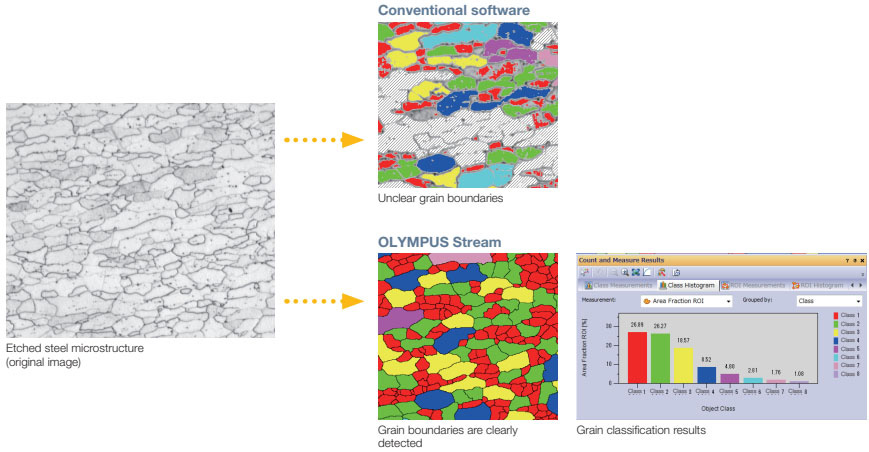
Particle Analysis вҖ“ Count and Measure Solution
Detecting objects and measuring size distribution are among the most important applications in digital imaging. OLYMPUS Stream softwareвҖҷs Count and Measure solution uses advanced threshold methods to reliably separate objects, such as particles and scratches, from the background. More than 50 different object measurement and classification parameters are available including shape, size, position, and pixel properties
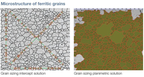
Grain Sizing in a Microstructure
Users can measure the grain size and analyze the microstructure of aluminum, steel crystal structures, such as ferrite and austenite, and other metals.
Supported standards: ISO, GOST, ASTM, DIN, JIS, GB/T
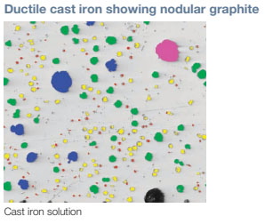
Evaluating Graphite Nodularity
The software can be used to evaluate the graphite nodularity and content in cast iron samples (nodular and vermicular). The form, distribution, and size of graphite nodes can be classified.
Supported standards: ISO, NF, ASTM, KS, JIS, GB/T
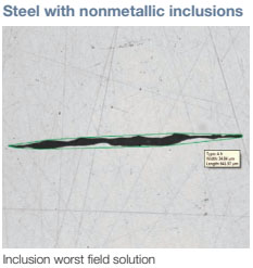
Rating Nonmetallic Inclusion Content in High-Purity Steel
Classify nonmetallic inclusions using an captured image of the worst field or inclusion that you have manually located on the sample.
Supported standards: ISO, EN, ASTM, DIN, JIS, GB/T, UNI
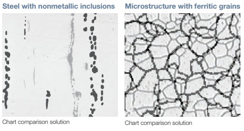
Compare Images of Your Sample and Reference Images
Easily compare live or still images with auto-scaled reference images. This solution includes reference images in accordance with various standards. The solution also supports multiple modes, including live overlay display and side-by-side comparison. Additional reference images can be purchased separately.
Supported standards: ISO, EN, ASTM, DIN, SEP
Material Solution Specifications*
| Solutions |
Supported standards |
| Grain intercept |
SO 643: 2012, JIS G 0551: 2013, JIS G 0552: 1998, ASTM E112: 2013, DIN 50601: 1985, GOST 5639: 1982, GB/T 6394: 2002 |
| Grain planimetric |
ISO 643: 2012, JIS G 0551: 2013, JIS G 0552: 1998, ASTM E112: 2013, DIN 50601: 1985, GOST 5639: 1982, GB/T 6394: 2002 |
| Cast iron |
ISO 945-1: 2010, ISO 16112: 2017, JIS G 5502: 2001, JIS G 5505: 2013, ASTM A247: 16a, ASTM E2567: 16a, NF A04-197: 2004, GB/T 9441: 2009, KS D 4302: 2006 |
| Inclusion worst field |
ISO 4967 (method A): 2013, JIS G 0555 (method A): 2003, ASTM E45 (method A): 2013, EN 10247 (methods P and M): 2007, DIN 50602 (method M): 1985, GB/T 10561 (method A): 2005, UNI 3244 (method M): 1980 |
| Chart comparison |
ISO 643: 1983, ISO 643: 2012, ISO 945: 2008, ASTM E 112: 2004, EN 10247: 2007, DIN 50602: 1985, ISO 4505: 1978, SEP 1572: 1971, SEP 1520: 1998 |
| Coating thickness |
EN 1071: 2002, VDI 3824: 2001 |
* Please see the OLYMPUS Stream brochure for more detailed information.
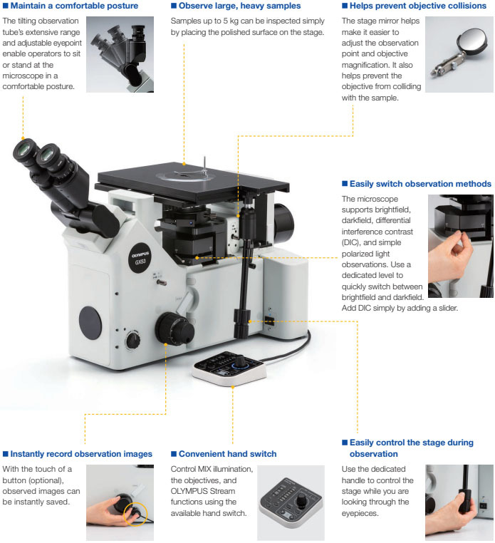
A Design That Emphasizes User Comfort
The microscopeвҖҷs ergonomic design helps users stay comfortable while they work, contributing to a more efficient inspection. When used with OLYMPUS Stream software, operators can easily acquire images of diverse samples, conduct a variety of analyses, and generate professional reports.
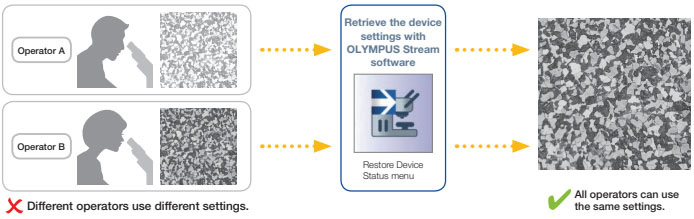
Easily Restore Microscope Settings: Coded Hardware
Coded functions integrate the microscopeвҖҷs hardware settings with OLYMPUS Stream image analysis software. The observation method, illumination intensity, and magnification can be recorded by the software and stored with the associated images. Since the settings can easily be reproduced, different operators can conduct the same quality inspections with limited training.
User Guidance Helps Simplify Advanced Analysis
The software guides users step-by-step through an inspection process that complies with the chosen industrial standard. Operators at any experience level can quickly and easily conduct advanced analysis by following the on-screen guidance.
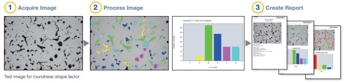
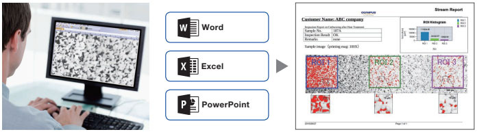
Efficient Report Generation
Creating a report can often take longer than capturing the image and taking the measurements. OLYMPUS Stream software provides intuitive report creation to repeatedly produce smart and sophisticated reports based on predefined templates. The software can be configured so that the magnification is printed along with individual images.
Proven optics and digital imaging technology deliver quality inspection data
OlympusвҖҷ history of developing high-quality optics and advanced imaging capabilities has led to quality microscopes that offer exceptional measurement accuracy.
Applications
Reflected light microscopy spans a range of applications and industries. Below are just a few examples of what can be achieved using different observation methods.
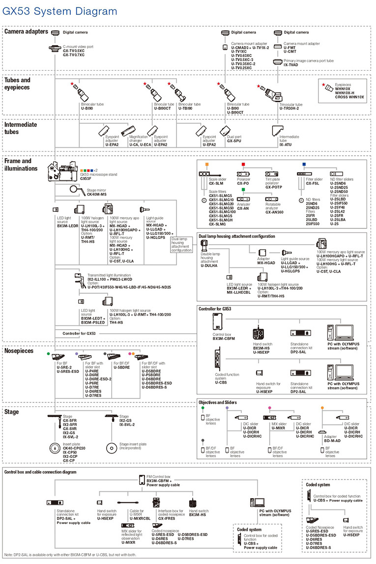
Choose the Components You Need
The GX53 microscope is designed to enable users to choose a variety of optical components to suit individual inspection and application requirements. The system can utilize all available observation methods. Users can also select from a variety of OLYMPUS Stream image analysis packages to meet image acquisition and analysis needs.
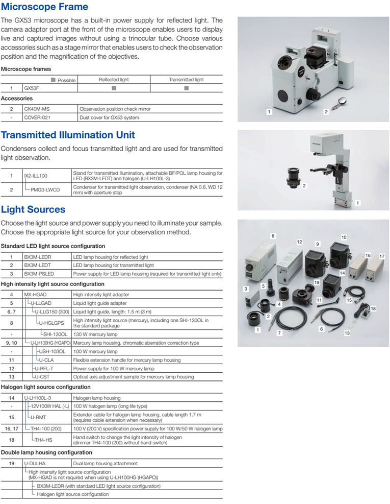
GX53 Reflected/Transmitted Light Combination
The GX53 microscope frame can be configured for both reflected and transmitted light with manual, coded, or motorized components.
Scales for Metallurgical Analysis
Glass scales can be inserted into the eyepiece to conduct observations that comply with industry standards. Grain size reticles, squared circles, and calibration scales are also available for each objective
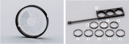
Scale slider
| 1 |
GX-SLM |
Scale slider, attachable 3 glass scales maximum |
| 2 |
GX51-SLMG5 |
Scale glass for 5Г— objective, scale length: 200 Ојm |
| 3 |
GX51-SLMG10 |
Scale glass for 10Г— objective, scale length: 100 Ојm |
| 4 |
GX51-SLMG20 |
Scale glass for 20Г— objective, scale length: 50 Ојm |
| 5 |
GX51-SLMG50 |
Scale glass for 50Г— objective, scale length: 10 Ојm |
| 6 |
GX51-SLMG100 |
Scale glass for 100Г—, scale length: 10 Ојm |
| 7 |
GX51-SLMGS |
Grain size scale, applied to JIS G 0551, ISO 643 and ASTM E112 AUSTENITE GRAINS IN STEEL PLATE IV No.1 to 8 |
| 8 |
GX51-SLMGH |
Lattice pattern, applied to JIS G 0555 |
| 9 |
SLMG |
Parfocal glass to adjust the light path length |
Build Your System Your Way

|