| |
 |
 |
home |
> |
м ңн’ҲмҶҢк°ң |
> |
кҙ‘н•ҷнҳ„лҜёкІҪ |
> |
мІӯм •лҸ„кІҖмӮ¬м„Ө비 |
> |
Inclusion Inspection system |
|
|
| |
| мІӯм •лҸ„кІҖмӮ¬м„Ө비 |
| Xcellence |
| |
| System Features |
 |
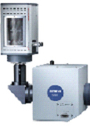
xcellence pro-for flexible live cell imaging
The Olympus xcellence proВ uses a sequential system coordinator board to provide dedicated control of all components as well as the MT10 highly stabilised illumination system. As a result, investigations planned using the Experiment Manager interface are followed precisely yielding excellent results. |
|
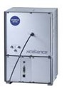 |
Olympus MT10
The MT10 is a highly stable illumination system, which incorporates a fast filter wheel (minimal switch 85 ms) with 8 positions, as well as a rapid attenuator with 7 illumination intensity grades. The built-in shutter opens and closes in less than 5 ms to reduce off-acquisition photobleaching. Two light source types are available: a 150W Xe arc burner or a 150W Hg/Xe mixed gas arc burner . The illumination source is connected to the microscope via a single quartz light fibre, while an electronic feed-back loop and a photo diode ensure perfect light intensity with minimal fluctuations. |
|
 |
xcellence rt-for precise real-time imaging
The Olympus xcellence rttakes experimentation to another level, employing a parallel real-time controller to ensure consistent component control and data uptake by the xcellence system during an experiment.В As a result, experiment control and precise sub-millisecond timings are maintained when changing hardware settings. The real-time controller communicates with all components of the MT20 illumination system and other peripherals in parallel ensuring seamless experiments, to provide reliable data as well as outstanding images. |
|
 |
Olympus MT20
The MT20 incorporates a fast, 8 position filter wheel (minimal switchВ 58 ms) and a fast attenuator providing 14 intensity grades. The built-in shutter has an exceptional 1 ms on/off time, thus eliminating off-acquisition photobleaching. Two reliable light source types are available: a 150W Xe arc burner and a 150W Hg/Xe mixed gas arc burner. An electronic feed-back loop and a photo diode ensure perfect light intensity with minimal fluctuations. The light is connected to the microscope via a single quartz light fibre, and every step is controlled with Вөs precision by the xcellence real-time controller. |
|
 |
Optical quality
xcellence fully controls all motorised modules of the Olympus IX2 and BX2-WI microscopes.В The optical bench concept and optical quality of these microscopes make them the perfect choice for advanced microscopy. |
|
 |
Cameras unlimited
In order to provide the right camera for any application, various monochrome CCD and intensifying EMCCD cameras, as well as colour cameras are supported by the xcellence system. Specialised cameras are provided for IR imaging that boast increased sensitivity in the IR range, allowing super fast imaging up to 500 fps and low light fluorescence imaging with sensitivity up to 98% Quantum Efficiency. Imaging speed can be increased by using binning and reducing the scanning area via the selection of specific regions of interest. Depending on the type of camera used, advanced camera functions such as overlapping readout, streaming, variable EM-gains, Real-gain and Photon-Imaging Modes can also be utilised. |
|
 |
Live cell imaging
Live cell imaging often means incubation, and different samples regularly need different environments. Olympus offers a broad range of environmental control systems, from simple heating plates, to bench top and cage incubators. Cooling, heating and the control of humidity and gas levels can be integrated into the xcellence imaging microscope systems.
|
|
 |
Intuitive graphical experiment planning, set-up and execution
The intuitive drag-and-drop functionality of the Experiment Manager enables the rapid assembly of complex experiments via icons that represent simple commands, groups of commands or entire sub-experiments. Individual parameters can be set for each image acquisition command and experiment plans are automatically stored together with the image data in the database. |
|
 |
Advanced software functionality
Alongside the Experiment Manager, xcellence also offers a range of advanced software functions including: image processing (e.g. Spectral Unmixing-a unique tool for colour resolution enhancement); a suite of image analysis tools; and a sophisticated, structured database for archiving and documentation.
|
|
 |
Functional modules
The xcellence systems are compatible with a large number of software modules and additional devices, making them the perfect choice for a wide range of experimental requirements. Different motorised stages can be used and controlled more quickly and precisely than when employing traditional imaging systems. There is also the option to combine the system with additional hardware, such as fast filter wheels, piezo Z-drives and the Olympus Zero Drift hardware autofocus. In addition, a bidirectional trigger interface allows synchronisation of up to sevenВ peripheral devices via TTL pulses.В В В В В В В
В |
|
| |
| Software Features |
|
Intuitive graphical experiment planning, set-up and execution
The intuitive drag-and-drop functionality of the Experiment Manager enables the rapid assembly of complex experiments via icons that represent simple commands, groups of commands or entire sub-experiments. Individual parameters can be set for each image acquisition command. Experiment plans are automatically stored together with image data in the database. |
|
 |
Time-lapse Imaging
Dynamic processes such as cell growth, metabolic transport and signal transduction are routinely monitored. The duration of such processes may vary from the sub-second range to hours or even days. Consequently it may be necessary to take several images per second or just one image every couple of minutes. The xcellence realtime system is a valuable tool, independent of whether you need up to 500 fps or just one frame per minute. It combines the fastest image acquisition possible for complex experiments with the interactivity you need to correct conditional changes over long experiments. |
|
 |
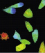
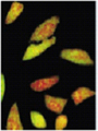
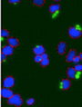
Multi-colour Imaging
The development of a growing list of specific fluorochromes covering the entire colour range enables the scientist to image and distinguish different sub-cellular structures simultaneously within one experiment, through the use of multiple staining. If this is combined with time-lapse acquisition, the illumination unit of the microscope must be able to switch quickly between excitation wavelengths. With the xcellence software, 3 clicks are enough to set up a new multi-colour experiment, start the acquisition and store the data in the database. |
|
 |
Z-sectioning and Multi-dimensional Imaging
Microscopy is fundamentally a 2D observation technique, while biological samples are inherently 3D. Therefore, in order to map the entire volume of the specimen it is necessary to construct an image from multiple layers. This can be achieved by moving the focal plane in precise steps using a motorised Z-drive or a piezo-electric drive. The xcellence system automatically calculates the perfect step distance and can be combined with an in-built piezo-stepper to provide the fastest and most accurate image acquisition possible, across a range of objectives.
|
|
 |
Ion Imaging / Ratio Imaging / Ca++ Imaging
The fluorescence behaviour of several dyes is dependent on the concentration of certain ions such as calcium (Fura-2) or on the pH value (BCECF). The detection, quantification and analysis of changes in fluorescence intensity are thus an indirect means to study important physiological processes. The xcellence software offers a versatile tool for ratio measurement, as well as the calculation of ion concentrations. To accelerate workflow, the xcellence system provides graphs and ratio images in real-time during image acquisition.
Read the Application note about Calcium signal transduction and mitochondrial dynamics hereВ |
|
 |
Spectral unmixing: enhanced colour resolution
Pronounced spectral overlap of the excitation and emission characteristics often prevents the combination of certain fluorescent markers within one sample. Linear spectral unmixing ascertains the contribution of different fluorochromes to the total signal and, by chromatic redistribution of the intensity, restores a clear signal for each colour channel, undisturbed by the emission from other fluorochromes. This procedure uses the inherent information present within the data, and the results are therefore not simply embellished images. The spectral redistribution maintains the overall intensity of the image and facilitates quantitative analyses, for example in co-localization and FRET studies. |
|
 |
Bleaching Correction
The xcellence software can be used to apply bleaching correction algorithms across a time series to ensure that during long term imaging, the effect of any unwanted bleaching is corrected. The correction parameters can be applied across the entire image or specific ROIs. |
|
 |
Kymograph
The advanced kymograph feature in the xcellence software enables 2D visualisation of inherently 3D events such as vesicle movement. Velocities and speed changes can be directly inferred via the kymograph image. xcellence offers precise calculation of the speed of molecule movement within a cell, even along convoluted pathways. |
|
 |
Measurements
xcellence features a unique working environment suitable for interactive measurement tasks and dimension calculations. Results can be presented as graphs or spreadsheets and evaluated directly to determine, amongst others, mean values, extremes and standard deviations. Making analysis simpler and faster, the Image Statistics button provides results from the 10 most popular statistical tests, both for the image and all defined ROIs, all in one easy to manage table. All measurements can be exported to Excel with just one click. |
|
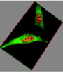 |
Image processing and 3D visualisation
The xcellence software provides a host of processing functions such as background subtraction, shading correction, display adjustment as well as filtering and overlay of fluorescence and transmission images. Deblurring algorithms (no neighbour, nearest neighbour and wiener filter) enhance the spatial resolution of widefield microscopy by reducing out-of-focus blur to yield images with greatly improved clarity. The SliceViewer generates slices through image stacks while the VoxelViewer renders 3D images and iso-surfaces.
|
|
 |
Fluorescence analysis and kinetics
More complex analysis routines are possible using the system, such as measurement of intensity kinetics, co-localization statistics as well as ratio and О”F/F analyses. Results are provided as false-colour images, graphs of regions of interest and data sheets.
|
|
 |
Programming
The programming language ‘Imaging C’ offers advanced users a complete, integrated development environment. This includes a programming library with unrestricted access to the wide range of image acquisition and processing functions. ‘Imaging C’ consists of a compiler, an interpreter, a macro recorder, a multiple-document (MDI) text editor, a symbol and function browser, and an interactive debugger with single-step processing, conditional breakpoints as well as a browser for variables and constants. |
|
 |
Archiving
The integrated database archives multi-dimensional image series along with analytical data, reports and associated documents in a well-structured system. The database has been designed to facilitate fast access and easy navigation, while keeping network capacity requirements low. |
|
 |
Report generation
The report generator makes creating reports that are compliant with various standards quick and easy via a simple drag-and-drop process. These reports can contain graphical elements such as images, sheets and diagrams and also offer a means of uncomplicated text input. Placeholders are automatically filled with the contents of various fields from information in the archive or from analytical results. |
|
| |
| Functional Modules |
 |
Xride 3D Deconvolution
The new XRide 3D deconvolution algorithm included in the xcellence software, provides faster and more accurate deconvolution, while ensuring outstanding usability. Faster than other algorithms (e.g. no neighbor, nearest neighbor, Wiener etc) and yielding near confocal results, the Olympus xcellence software provides a new level of precision and speed to deconvolution. |
|
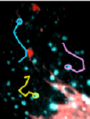 |
trackIT!
A number of biological processes require molecules to move into, out of and within cells. Tracking this movement can provide key insights into the biology and the biochemistry of these movements. The TrackIT feature automatically detects moving objects and tracks them over a given period of time. Additionally, semi-automatic tracking enables the user to track these events by hand and let the software do all the rest.В The results are presented as movies showing object tracks, accompanied by charts and histograms of parameters such as velocity, direction, path length and total distance. |
|
 |
FRET (Foerster Resonance Energy Transfer) analysis
The measurement of fluorescence energy transfer from one fluorochrome molecule to an adjacent one can be used for the investigation of molecular interactions in cells. It requires the acquisition of images with different excitation and emission wavelengths and sophisticated correction algorithms.В The FRET measurement package is easy to use and very flexible offering both predefined or custom algorithms. |
|
 |
Multidimensional Object Detection
The Multidimensional Object Detection tool analyses 4D image data. Objects can be detected and classified based on diverse parameters such as area, size, shape, position, density and intensity. The Multidimensional Object Detection tool has multiple applications including automatic cell classification, cell counting, mitotic- and apoptotic analysis, drug discovery and many more. |
|
 |
Alternative illumination systems
To provide speed and flexibility, the xcellence systems can integrate with specialised light-sources such as the multi-colour LED illumination system precisExcite and the Monochromator Poly 5000. These systems offer switching times of less than 5 ms and are controlled with Вөs precision by the xcellence rt real-time controller board. |
|
 |
The new ZDC2: z drift compensation in real-time
During long time-lapse observations, temperature changes can cause focal drift, resulting in a change of focal plane. This can be a serious problem, for example in TIRF microscopy, because of the high z-resolution required (100nm). The Olympus ZDC2 (Z Drift Compensation) automatically corrects for thermal drift prior to imaging. Compensation is performed using a 785nm IR laser, which detects the bottom surface of the experimental dish with exact precision. |
|
 |
High speed multi-dimensional acquisition
The xcellence system offers multiple solutions to speed up image acquisition. The PIFOC®  microscope NanoPositioner has a piezo-based z-drive built into the microscope frame to increase the speed and accuracy of z-stack acquisition. To speed up multi-colour imaging, a fast fluorescence turret (300ms), observation filter wheel (58ms) and shutter (37ms) can be used. |
|
 |
Simultaneous multi-colour imaging
The DV2 Dual-Viewв„ў Micro-Imager beam-splitting device can be added to allow the simultaneous acquisition of two chromatically separated fluorescence images. With the Quad-Viewв„ў system, up to 4 colours can be separated and simultaneously acquired with the xcellence software. For simultaneous imaging of the full field of view the xcellence system is capable of synchronized acquisition with two cameras using the DC2 Dual-CamTM. |
|
 |
Structured illumination
The OptiGrid structured illumination system is capable of removing out of focus light thereby creating results similar to those achieved by confocal microscopy. The system can be fully integrated with xcellence to produce ultra-rich, multi-channel fluorescence 3D/4D images using standard illumination at a range of wavelengths, from UV to IR. Furthermore, the OptiGrid MВ works at a market-leading speed and is capable of acquiring many frames per second. |
|
 |
Confocal Spinning Disk
The DSU (Disk Scanning Unit) offers very fast and cost efficient confocal image capture via a cooled CCD camera and the MT10/20 illumination system. The DSU is the perfect application for avoiding photo-bleaching and photo toxic effects, which are problems that often influence fluorescence live cell imaging.В The DSU disk contains a pattern of slits that creates a virtual pinhole as the disk spins. By removing out of focus blur, resolution is enhanced, yielding clear, continuous, optically cross-sectioned images. |
|
 |
cell^tirf
Total internal reflection fluorescence microscopy (TIRFM) has firmly established itself over the last few years as a key technique in the investigation of molecular interactions at or near the cell surface. Olympus is experienced in providing advanced TIRFM solutions and cell^tirf takes this to the next level with a series of peerless features. |
|
 |
cell^frap
The Olympus xcellence cell^frap providesВ an advanced system for photo-manipulation applications including: fluorescence recovery after photo-bleaching (FRAP), inverse FRAP (iFRAP), fluorescence loss in photobleaching (FLIP) and fluorescence loss after photobleaching (FLAP). The cell^frap application is also ideal for more complex processes such as photo-conversion, photo-activation, pattern bleaching, intra cellular laser cutting and laser trapping.
|
|
 |
Additional photo-manipulation devices
The FLASH/C2 is a Pulsed Xenon Light Source for Photolysis and Bleaching. Regions of different sizes can be easily stimulated with 0.6-1ms pulses. For increased power and wavelength flexibility, the laser based Point-FRAP system can be used, which restricts illumination to a refraction limited spot that can be manually selected via the field of view. Both systems can be used in real time with the xcellence system. |
|
| |
| Specifications of xcellence pro |
| Hardware В В |
Olympus microscopes BX61WI, IX50-IX81, MVX10 |
|
| CCD, EMCCD cameras, various models |
|
| Imaging Computer (latest generation PC) |
|
| В |
В |
| Illumination system В В |
MT10 |
В |
| Short arc burners, |
150 W Xenon or Mercury-Xenon |
| 8 Filter positions |
diameter 25 mm |
| Filter switch |
min. 85 ms (neighbouring positions) |
| Attenuation |
7 levels, 4% - 100% |
| Attenuation switch |
В <85 ms |
| Shutter, on/off time |
5 ms |
| Operation |
sequentially |
| The xcellence system can be also equipped with other illumination systems like Poly 5000, precisExcite LEDВ |
В |
| В В В В |
В В В |
| Hardware control В |
Control board with additional CPU, independent from imaging PC |
System coordinator |
| Temporal resolution |
10 ms (camera 1 ms) |
| Timing precision |
ca. 15 ms |
| Camera control |
trigger (level trigger if accepted by camera) |
| Peripheral device control via TTL pulses |
optional:В 7 BNC connectors |
| Multi-task acquisition with hardware switch (z-position, exciter filter etc.), 50 ms exposure |
3-4 full frames/sec with sequential hardware switch |
| В |
|
| Experiment set-up and control В |
Graphical interface Experiment Manager |
|
| Drag-and-drop alignment of command icons |
|
| 7-D acquisition (xyz, excitation and emission colour, time, stage position) |
|
| Loop-in-loop capability (repetition of complex command groups) |
|
| Experiments with varying acquisition speed and camera exposure |
|
| Autofocus, repeatedly during experiment |
|
| Live image display and online analysis |
|
| User interaction: pause, resume, set marker |
|
| В |
|
| Imaging software В |
xcellence proВ |
|
| Structured database for multi-dimensional data handling and storage |
|
| Image types: (n x 16) bit, 8 - 24 bit export and import |
|
| Image processing: filters, extended focal imaging, shading and background correction, arithmeticвҖҰ |
|
| Measurements and analyses: number, length, distance, area, circumference, angle, grey value, histograms, line profiles, tables, statistics, diagramsвҖҰ |
|
| Fluorescence analyses: intensity kinetics, ratioing, О” F/F, kymograph |
|
| Spectral unmixing for optimised colour resolution |
|
| Deblurring, Bleaching Corrrection, SliceViewer, VoxelViewer |
|
| Macros and automated functions: imaging C module, macro recorder |
|
| В |
|
| Options В В В |
Piezo-electric objective drive (all microscopes) or nosepiece drive (IX51, IX71, IX81 only) |
|
| Fast observation filter wheel (60 ms) and fast motorized mirror unit turret |
|
| TIRFM lasers and illumination combiners, multi-line |
|
Motorised cell^tirf illuminator |
|
| cell^frap galvo-scanning photo-stimulation module |
|
| Point-FRAP System |
|
| DualView Image Splitter |
|
| Optigrid M structured illuminaton system |
|
| DSU spinning-disk-confocal |
|
| FRET, hardware and analysis (different algorithms) |
|
| Deconvolution for resolution enhancement |
|
| Automatic and semi-automatic object tracking |
|
| Particle detection and tracking |
|
| В |
|
| Upgradeability |
To xcellence rt |
|
|
| |
| Specifications of xcellence rt |
| Hardware В В В В В |
Olympus microscopes BX61WI, IX50-IX81, MVX10В |
|
| CCD, EMCCD cameras, various models |
|
| Imaging Computer (latest generation PC) |
|
| В |
В |
| Illumination system В В В В В В В В В |
MT20 |
В |
| Short arc burners, |
150 W Xenon or Mercury-Xenon |
| 8 Filter positions |
diameter 25 mm |
| Filter switch |
min. 58 ms (neighbouring positions) |
| Attenuation |
14 levels, 1% - 100% |
| Attenuation switch |
В <58 ms |
| Shutter, on/off time |
1 ms |
| Operation |
all modules in parallel |
| xcellence system can be also equipped with other illumination systems like Poly 5000, precisExite LED |
В |
| В |
В |
| Hardware control В В В В В В В |
Control board with additional CPU, independent from imaging PC |
Real-time Controller |
| Temporal resolution |
1 ms |
| Timing precision |
< 0.01 ms |
| Camera control |
trigger (level trigger if accepted by camera) |
| Peripheral device control via TTL pulses |
7В BNC connectors |
| Multi-task acquisition with hardware switch (z-position, exciter filter etc.), 50 ms exposure |
8 full frames/sec with parallel hardware switch |
| В |
В |
| Experiment set-up and control В В В В В В В В |
Graphical interface Experiment Manager |
|
| Drag-and-drop alignment of command icons |
|
| 7-D acquisition (xyz, excitation and emission colour, time, stage position) |
|
| Loop-in-loop capability (repetition of complex command groups) |
|
| Experiments with varying acquisition speed and camera exposure |
|
| Autofocus, repeatedly during experiment |
|
| Live image display and online analysis |
|
| User interaction: pause, resume, set marker |
|
| В |
|
| Imaging software В В В В В В В В В |
xcellence rt |
|
| Structured database for multi-dimensional data handling and storage |
|
| Image types: (n x 16) bit, 8 - 24 bit export and import |
|
| Image processing: filters, extended focal imaging, shading and background correction, arithmeticвҖҰ |
|
| Measurements and analyses: number, length, distance, area, circumference, angle, grey value, histograms, line profiles, tables, statistics, diagramsвҖҰ |
|
| Fluorescence analyses: intensity kinetics, ratioing, О” F/F, kymograph |
|
| Spectral unmixing for optimised colour resolution |
|
| Deblurring, Bleaching Corrrection, SliceViewer, VoxelViewer |
|
| Macros and automated functions: imaging C module, macro recorder |
|
| В |
|
| Options В В В В В В В В В В В В |
Piezo-electric objective drive (all microscopes) or nosepiece drive (IX51, IX71, IX81 only) |
|
| Fast observation filter wheel (60 ms) and fast motorized mirror unit turret |
|
| TIRFM lasers and illumination combiners, multi-line |
|
Motorised cell^tirf illuminator |
|
| cell^frap galvo-scanning photo-stimulation module |
|
| Point-FRAP System |
|
| DualView Image Splitter |
|
| Optigrid M structured illuminaton system |
|
| DSU spinning-disk-confocal |
|
| FRET, hardware and analysis (different algorithms) |
|
| Deconvolution for resolution enhancement |
|
| Automatic and semi-automatic object tracking |
|
| Particle detection and tracking |
|
|
| |
|
| |
|