| |
 |
 |
home |
> |
м ңн’ҲмҶҢк°ң |
> |
м „мһҗнҳ„лҜёкІҪ |
> |
мЈјмӮ¬м „мһҗнҳ„лҜёкІҪ(SEM) |
> |
JASM-6200 ClairScopeв„ў |
|
|
| |
| мЈјмӮ¬м „мһҗнҳ„лҜёкІҪ(SEM) - JEOL |
| JASM-6200 ClairScopeв„ў |
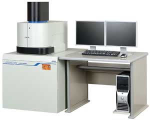
| JASM-6200 Scanning Electron Microscope ClairScopeв„ў |
A Scanning Electron Microscope capable of sample imaging in atmosphere
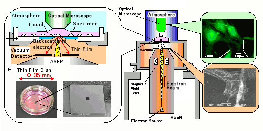
Design Principle
ClairScopeв„ў consists of an atmospheric scanning electron microscope (ASEM) and an optical microscope positioned on top. The ASEM is isolated from the SEM with a thin film installed at the top end of the inverted SEM. The thin film, designed to transmit an electron beam while blocking air, separates the sample in atmosphere from the vacuum in the SEM. The electron beam is projected from below to the sample placed on the thin film for high resolution imaging of the sample in atmosphere. The same area of view of the sample can also be imaged in the optical microscope on top. The thin film is configured in a dish (thin film dish), which can be used for cell culture in a culture chamber.
|
 |
|
 |
Features
- SEM imaging in complete atmospheric pressure
The sample is held in complete atmospheric pressure, enabling dynamic observation of physical and chemical reactions in liquids and gases. Lengthy preliminary treatment of biological samples, including dehydration and drying, is no longer necessary, resulting in high throughput imaging. The microscope, without the limitations of vacuum atmosphere, will broaden the range of applications.
- Imaging of the same area of view as optical microscope
The top of the thin film is open, joined to the optical microscope. The optical microscope being aligned with the ASEM, the operator can image the same area of view alternately between both microscopes.
The optical microscope can accommodate up to 6 mirror units simultaneously. Standard mirror units for bright field imaging and ultraviolet irradiation, combined with optional mirror units, can acquire various types of fluorescent images.
- Open specimen chamber
The specimen chamber is open, allowing the operator to externally control reagents (chemical administration).For example, the operator can load the thin film dish onto the system, and observe the sample in the ASEM after administering a chemical to the sample. The operator can also monitor physical and chemical developments as the sample changes its volume.
|
|
Biological applications
- Extended applications of optical microscope
Many biologists routinely use optical microscopes for their research. However, optical microscopy is unable to achieve resolution higher than 0.2 um due to its wavelength limitations. While scanning electron microscopy (SEM) and transmission electron microscopy (TEM) feature higher resolution, many samples require a lengthy preliminary treatment process including multiple dehydration and drying process steps, which usually takes a skilled technician one to a few days to complete. This is why SEM and TEM, despite their high resolution, are not as widely used as optical microscopy.
ClairScopeв„ў is designed to observe samples in atmospheric pressure using the ASEM. It is capable of high resolution imaging of biological samples without the preliminary dehydration/drying process that requires skilled technician. Sample pre-treatment consists of simple steps of chemical administration, taking only about 10 minutes. This enhances the efficiency and yield of sample imaging.
ClairScopeв„ў also supports imaging of the same area of view in the optical microscope on top and the ASEM at the bottom. This allows optical microscope users to first observe images they are familiar with, and proceed to high resolution imaging of a given spot of the sample. The system allows the operator to identify tissues and local existence of protein using fluorescence staining and optical microscopy, and to further study the corresponding spots using high resolution imaging of the ASEM.
ClairScopeв„ў also allows the operator to observe live cells in the optical microscope, restrict the motion with chemicals, and apply fixation/staining as needed for high resolution imaging in the ASEM. Cells are cultured on the thin film dish in the same way as any conventional plastic dish. The researcher can culture cells in the usual procedure, observe the cells in the optical microscope, and focus on areas of interests in the ASEM for further imaging.
ClairScopeв„ў is expected to be widely used in a variety of fields including basic biology, medicine, pharmaceutical, and cosmetics.
|
|
Other applications
- Dynamic observation of chemical reaction
Real time high resolution imaging can reveal the mechanism of chemical reactions in liquid and vapor. Traditionally, this has been accomplished by TEM with an environmental cell. However, TEM can only accept thin film samples, and SEM imaging has been in demand for certain samples. ClairScopeв„ў, designed to image samples in atmospheric pressure in the ASEM, is capable of real time imaging of chemical reactions in liquid and vapor. The open top of the thin film facilitates chemical administration, and enables observation of gas emitting reactions. Furthermore, the system allows the optical microscope on top to alternately observe the same area of view as the ASEM. The system has succeeded in dynamic observation of hydrolytic reactions of plaster.
- Dynamic observation of drying process
Drying is a critical process in various industries. However, it has been difficult to achieve dynamic observation of the drying process at higher resolution than optical microscopy. ClairScopeв„ў, with the open top of the thin dish, enable observation of the drying process of solvents. It has succeeded in dynamic observation of the drying process of a salt containing solvent from drying to crystallization.
- Dynamic observation of electrochemical reaction
Electrochemical reactions, related to batteries, plating, corrosion, and refinement, is essential in many industries. High resolution imaging of electrochemical reactions in liquid has been a difficult issue. ClairScopeв„ў, capable of real time high resolution imaging of reactions in liquid in atmospheric pressure in the ASEM, also supports dynamic observation of electrochemical reactions. With the open top of the dish, the system is also effective in observing electrochemical reactions generating gases. It has succeeded in dynamic observation of electrochemical reactions of a sample loaded onto a prototype thin film dish incorporating an electrode in the thin film.
- Basic physics
Particles in liquid are known to demonstrate Brownian motion and self assembly under certain conditions. These reactions are related to the basic motion mechanism of life, a field to be explored extensively. These reactions have been studied in optical microscopy. ClairScopeв„ў has succeeded in real time observation of Brownian motion and self assembly of particles.
- Reaction to electron beam irradiation
A high intensity electron beam can induce reactions in liquid and vapor. Using ClairScopeв„ў, the operator can induce such reaction in the ASEM for dynamic imaging, and also monitor the reaction using the optical microscope on top.
|
ClairScopeв„ў JASM-6200 Specifications
|
|
| Atmospheric scanning electron microscope (ASEM) |
| Resolution |
8 nm (30 kV) |
| Magnification |
100 to 100,000x |
| Accelerating voltage |
10, 20, 30 kV |
| Optical microscope (OM) |
| Mirror unit |
Up to 6 (bright field imaging and ultraviolet irradiation as standard) |
| Objective lens |
40x liquid immersion lens without cover |
| CCD camera |
High sensitivity color CCD, 1,600 x 1,200 pixels |
| Sample holder |
| Thin film dish |
35 mm dia; window 0.25 mm square (volume approx. 10 cc) |
| Other |
| Computer |
IBM PC/AT compatible |
| OS |
WindowsВ® Vista* |
| Viewing monitor |
2 19вҖқLCD units |
|
| |
|
|
|
|